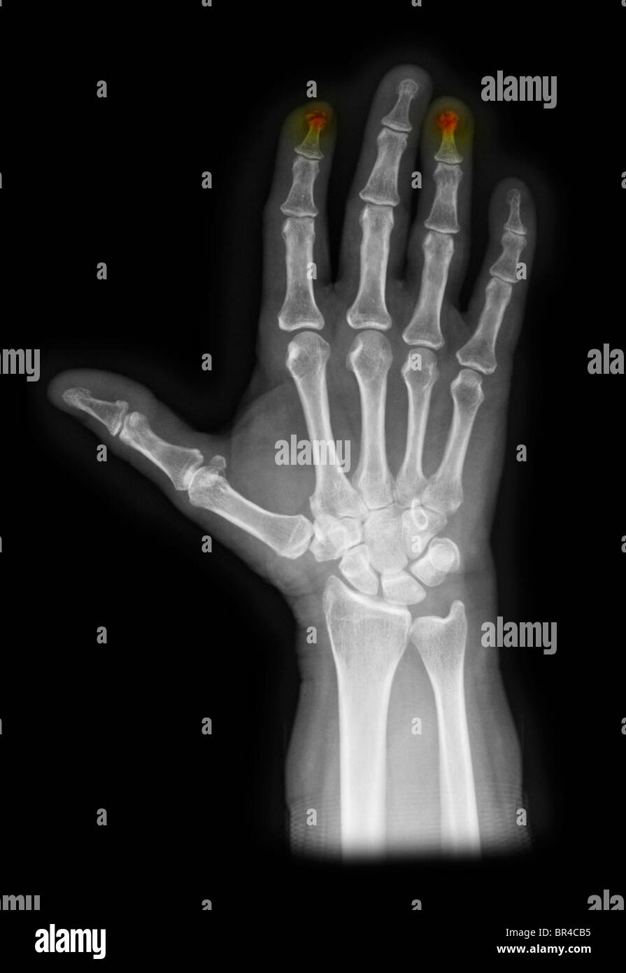
Key aspects of the history and physical are discussed, as well as additional testing that should be obtained.

The column begins with the reason and assesses questions that might go through a resident's mind as he or she heads to the emergency department to see the patient. Editor's Note: "Consult Corner" addresses a consult commonly encountered by an on-call resident. Specific operative treatment complications: Superficial and deep infection, deformity of the nail, secondary displacement of the fracture, joint incongruity, avascular necrosis, or extensor tendon rupture have been reported with operative management. The deformity is maintained by the contracture of the triangular ligament. Swan neck deformities: this occurs due to weakness of the volar plate and transverse retinacular ligament at PIPJ level, and dorsal slippage of the lateral bands with subsequent PIPJ hyperextension.
#Jammed finger xray full
There are no reported differences in the outcomes in terms of extensor lag or patient satisfaction following nonoperative treatment regardless of nighttime splinting after full time splinting. However, no functional deficits or patient dissatisfaction have been reported. Mild extensor lag < 10° and prominent bump on the dorsal aspect of the DIPJ: may be present following nonoperative or operative treatment.
#Jammed finger xray skin
A crucial part of the treatment is patient education on skin hygiene care without allowing DIPJ flexion.ĭorsal skin complications: These are the most commonly encountered complications e.g ulceration, maceration, nail deformity. It should be encouraged to keep the splint on at all times as removal and flexion of the joint reset the 6 to 8-week clock back to time zero.
#Jammed finger xray free
The consensus for extension splinting duration is 6-8 weeks, with progressive flexion exercises at six weeks. The PIPJ should be allowed a free range of motion with DIPJ only splinted in extension or slight hyperextension. The finger should remain splinted until seen by a hand specialist. Perforated splints have better compliance than the traditional solid splints. Yet, the type of splint, the duration of full-time wear, and the need for supplemental night orthotic wear are typically based on the provider’s preference.Ĭonservative options include Stack splint, thermoplastic splint, or aluminum foam splint, all to achieve the same principle which is an extension or slight hyperextension at the DIP joint. For soft tissue mallet finger, acute and chronic, splints have been reported to be safe and highly effective. Although somewhat controversial, there is some consensus in the literature that in the absence of large articular surface disruption or subluxation, non-operative treatment with the placement of a splint is favorable for both soft tissue and bony mallet.

Ī number of treatments have been tried, ranging from reassurance to conservative splint placements to surgically corrective procedures. Mallet finger injuries result when the extensor tendon is disrupted. The tendons of the digits extend over three joints. The tendons on the top of the hand are called extensor tendons and extend or straighten the digits, while the flexor tendons on the pals side of the hand flex or bend the fingers. The muscles that move the digits (fingers and thumbs) are located in the forearm and are connected to the bones of the digits by long tendons. Tendons are tissues that connect muscles to bone. The joints sit in volar plates, which are collateral ligaments attached to dense fibrous connective tissue, to increase stability. These joints are the distal interphalangeal joint (DIPJ) the metacarpophalangeal joint (MCPJ), which is the joint that connects the digit to the carpal or hand bones, and the proximal interphalangeal joint (PIPJ), which is the joint between the DIP and MCP joint. They include two bones in the thumb or three bones in the fingers as well as two to three joints between the phalanges. The bones making up the digits are called phalanxes or phalanges. This activity reviews the pathophysiology of mallet finger and highlights the role of the interprofessional team in the management of patients with this condition. Though some athletes and coaches often believe mallet injuries to be minor, each case should have a systematic evaluation performed. Although it is the most common closed tendon injury seen in athletes as a result of high velocity and contact sports, it also can be the result of a relatively minor trauma such as doing household chores (tucking in a shirt, tucking in sheets) or work-related activities. As some individuals do not see the hammer resemblance, some have proposed changing the name to drop the word "finger" due to its appearance. Mallet, which means hammer, was the term used to describe the hammer-like deformity that occurred in sports-related injuries in the 1800s.

Mallet finger injuries are commonly encountered in everyday clinical practice.


 0 kommentar(er)
